Showing posts with label Digestion and Ingestion system. Show all posts
Published May 21, 2023 by

Digestive System: Definition, Anatomy and Functions


What is the digestive system?

The digestive system is known as the set of organs that are in charge of the digestive process, that is, the transformation of food so that it can be absorbed and used by all the cells of the organism.  In addition to humans, most of the higher animals have a digestive system that performs this function.

During digestion or the digestive process, the different types of nutrients found in the food consumed (carbohydrates, lipids and proteins) are transformed into simpler units, thanks to the different digestive enzymes.  Under these conditions, the most elementary usable parts of the nutrients can be absorbed and then transported through the blood to all the cells of the body, where they are used to obtain energy and carry out all the essential functions for the maintenance and development of life. .

The functional process of the digestive system includes all the events that take place, from the entry of food into the mouth, to the expulsion of feces (indigestible remains) through the anus, passing through the absorption of nutrients through the intestinal walls.  It is a long process, which involves a set of complex mechanisms, in which numerous organs and parts of the body are involved, and which is essential for life, given that human beings (like all animals) are heterotrophs, and therefore Therefore we can only incorporate the organic matter we need through food.


Digestive system anatomy

Food gives the body the energy necessary for life.  The digestive system processes food separating the basic nutrients that can be assimilated by the body. From the mouth, through the stomach and intestines, to the anus, lies the digestive system, which is a long, muscular tube. Food is digested as it passes through the digestive system, which transforms it into elements that can be taken into the circulation. Certain organs (such as the liver, gallbladder, and pancreas) help with digestion.  The parts of the food that cannot be digested are expelled from the body in the form of excrement.


Digestive tube

It extends from the mouth (entrance orifice) to the anus (terminal or exit orifice).  It measures about ten meters in length and is made up of several differentiated portions.


Mouth

It forms the entrance to the digestive tract.  It is in charge of receiving the food that is ingested and crushing it.  Three main functions are developed: chewing, insalivation and swallowing.


Pharynx 

Musculo-membranous cavity that is located at the back of the mouth and communicates at the top with the nasal cavity and middle ear, and at the bottom with the larynx and esophagus.  It has a double function: passage of breathed air and ingested food.


Esophagus 

Tube about 25 cm long that connects the pharynx with the stomach. It crosses the diaphragm and is located between the trachea and the spinal column.  Its main function is to carry food to the stomach.


Stomach

Hollow "J" shaped organ measuring approximately 25 cm in length, situated below the diaphragm.  Two orifices or valves are differentiated: cardia, which communicates with the esophagus, and pylorus, which serves as a limit with the small intestine.  The gastric mucosa that covers it is of special importance to protect it from the acids that are part of the composition of the gastric juice. In the stomach, food is crushed by mechanical agitation and unites with the gastric juice, forming a highly acidic mush called chyme.
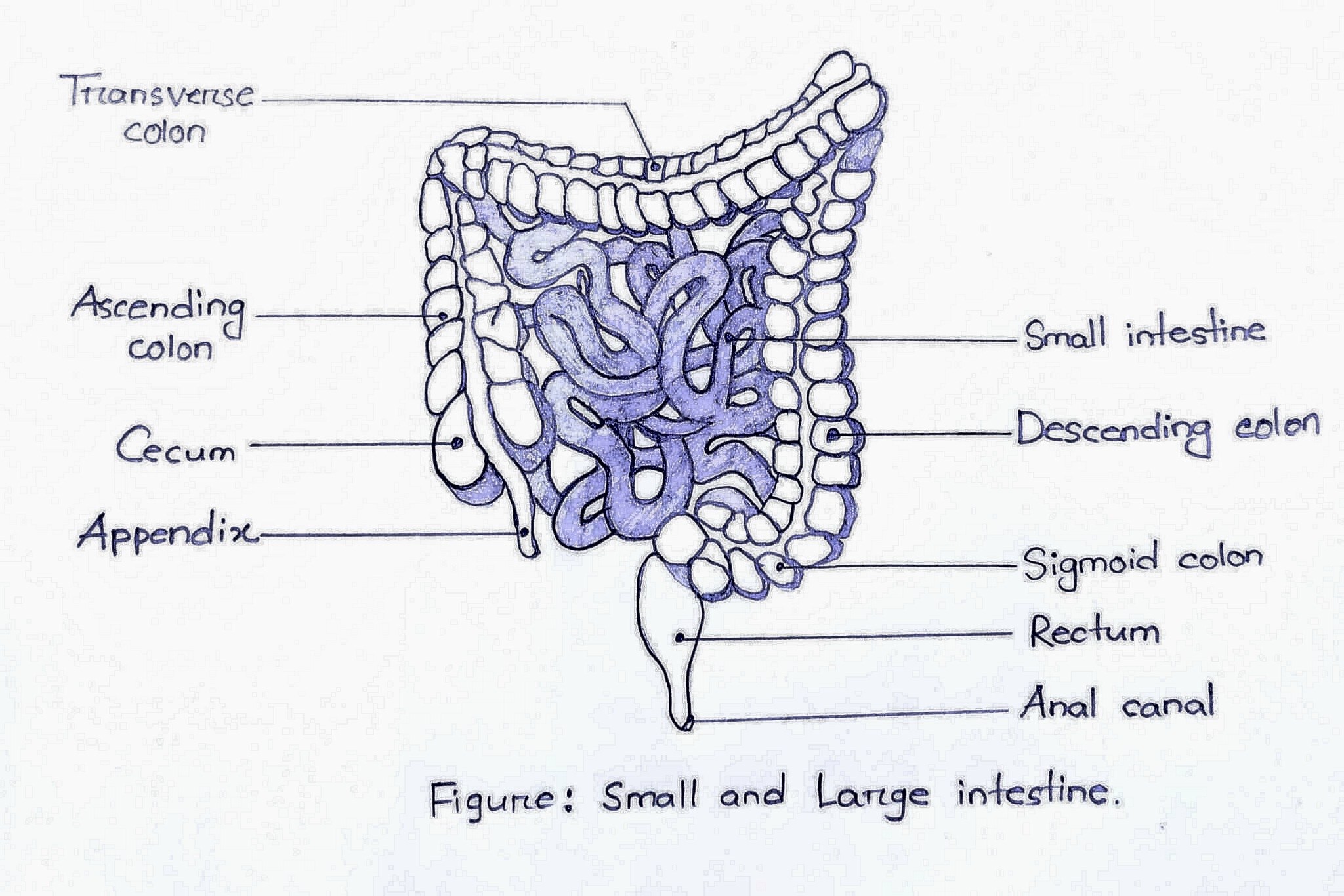

Small intestine  

Tubular structure situated below the stomach.  Under normal conditions it can measure between 6 and 7 meters in length and about 3 cm in width.  It occupies most of the abdominal cavity, where it remains rolled up.  Duodenum, jejunum and ileum are three major parts of small intestine.


Large intestine  

It is 1.5 to 2 meters long and extends from the ileocecal valve (which closes the opening of the small intestine) to the anus.  It consists of three segments: blind, colon and rectum, and communicates with the outside through the anus.  Its functions are the absorption of water and minerals and the elimination of waste products through the anal sphincter.


Function of various  parts of digestive system :

The basic function of the digestive system is the transfer of nutrients from the external environment to the internal environment, so that the cells of the organism have molecules that allow their metabolic maintenance and restoration.

The nutrients accessible to the organism can be of very different nature and size.  Those that are small in size can be absorbed without undergoing any change;  while those that have a large size have to go through a fractioning or breaking process that is called digestion.

The digestive system is not an excretory system, since most of the products that come out of it, the feces, correspond to non-absorbed material, bacteria and degraded cells of the digestive system itself.


Mouth

Chewing triggers the start of the digestive process in the mouth. 

The salivary glands produce saliva, a digestive juice that moistens food for easier transport down the esophagus to the stomach.


Pharynx 

The function of the pharynx is therefore to direct air or food to its proper place.  The oropharynx is part of the upper digestive tract, although it also serves as a passage for air in mouth breathing or coughing.  It goes from the pharyngeal entrance to the base of the epiglottis.  When we eat, we introduce food through the oral cavity where we chew it and then push it back with our tongue.  That "back" is the pharynx, a conduit where the already crushed and moist food can continue its process in the body until it reaches the stomach.  The pharynx is where the muscular contractions are carried out that will allow the food to continue its course and reach the esophagus.

The contractions also prevent food from entering the trachea and thus from leaking into the respiratory canal.  The epiglottis is the valve that, with contractions, the pharynx is in charge of closing and preventing us from choking on a piece of food.


Esophagus 

The esophagus is the initial part of the digestive tract and its function is to transport the food bolus from the pharynx to the stomach, through the thorax and prevent its reflux.


Stomach

Functions of stomach includes:

* temporarily store food

* mechanically break food into small particles

* mix the food bolus with the gastric secretion until obtaining a semi-liquid mass called chyme

* chemically digest proteins

* progressive emptying of chyme at a rate compatible with digestion and absorption by the small intestine

* secrete intrinsic factor, which is essential for the absorption of vitamin B12, into the ileum


Small intestine :

Long tube-shaped structure that joins the large intestine to the stomach. Its job is to keep the stomach's digestion going while absorbing nutrients (vitamins, minerals, carbs, lipids, and proteins) and water for the body to use. 


Large intestine

The following activities of the large intestine are all necessary for the body to function properly and include:

* Water absorption and stool formation

* Vitamins, short-chain fatty acids and nutrient recycling

* pH regulation

 *immune function

* function of the appendix


Read More
Published May 16, 2023 by

Anatomy, Histology and Function of Large Intestine

General Discussion  

A component of the digestive tract is the intestine, a long, winding tube.  The intestine aids in the extraction and absorption of nutrients and water from meals as well as the removal of waste.

The intestine has two main parts:
Food enters the small intestine from the lower portion of the stomach and is further digested there before moving on to the big intestine. The small intestine is about 20 feet (6.1 m) long.
The large intestine absorbs water, nutrients, and compounds (such salt and chloride) from digested food.  Additionally, it gets the body ready to expel waste (feces).  The term "colon" also refers to the big intestine.  It is roughly 5 feet (1.5 meters) long.

Anatomy
The terminal portion of the intestine extends from the ileocecal valve to the anus, although this valve does not properly correspond to the extremity of the large intestine.  Along this path, the large intestine measures 1.5 m in length, and its diameter is greatest at its origin, that is, in the ascending colon, where it measures 7 cm;  It narrows in the transverse and descending colon, where it reaches 4 cm, to dilate again when it reaches the rectum, giving rise to the rectal ampulla. The right colon begins at the cecum, continues with the ascending colon, continues at the hepatic flexure, and ends at the proximal transverse colon.  The splenic flexure, descending colon, sigmoid, rectosigmoid, and anal canal are all located in the left colon. The left colon also contains the distal portion of the transverse colon. The transverse and sigmoid colons are suspended in the peritoneal cavity by their mesenteries, while the ascending and descending colons are attached to the retroperitoneum.
The anal canal, which is located between the rectum above and the anal verge (anal orifice) in the perineum below, is the most terminal portion of the lower GI tract/large intestine.

Histological structure of the large intestine
The colon, rectum, and cecum make up the three sections of the large intestine.  Similar to the rest of the digestive system, the three sections of the wall have a similar structure.
It is made up of the four characteristic layers: the mucosa, submucosa, muscularis, and serosa.  The mucosa is supported by a band of fine bundles of smooth muscle (muscularis mucosa).
The morphology is quite similar between the three sections of the large intestine, so histological differentiation is not always possible.  In the large intestine there are no villi, the height of the mucosa is uniform.  This mucosa contains numerous deep and narrow crypts that span the full thickness of the mucosa.  Goblet cells predominate on the surface and entire length of the crypts.
There are muscular mucosa, and the submucosa is similar to the small intestine.  There may be lymphoid nodules developed in the submucosa and Meissner's nerve plexus.
Auerbach's nerve plexus is found between the circular and longitudinal muscle layer.

The serosa is composed of connective tissue and mesothelium.  In the final portion of the rectum the serosa becomes adventitious (lacks mesothelium)


Main functions of the large intestine
The large intestine performs the following functions, all of which are essential for the proper functioning of the body:
* Water absorption and stool formation
* Vitamins, short-chain fatty acids and nutrient     recycling
* pH regulation
*immune function
* function of the appendix

Water absorption and stool formation
A very important function of the large intestine is the absorption of water and the transformation of food material into feces, a process that takes place mainly in the colon.
By the time the chyme reaches the large intestine, most of the nutrients and 80-90% of the water have been absorbed.  At this point, electrolytes such as sodium, magnesium, and chlorine remain along with dietary fiber and other indigestible parts of food.
The ascending colon receives fecal matter still with a large amount of fluids.  The muscles of the colon are making peristaltic movements and move the fecal material slowly, the water is absorbed and the feces are acquiring more and more consistency until they reach the descending colon.
As it passes through the colon, the fecal material also compacts and mixes with mucus and bacteria from the natural flora of the intestine.  The mucus exerts a lubricating action to facilitate the advance of the feces, while the bacteria ferment the dietary fiber.

Vitamins, short-chain fatty acids and nutrient recycling
Bacterial metabolism ferments various types of dietary fiber and produces short-chain fatty acids, such as acetate, propionate, or butyrate.  These fatty acids are nutrients used by the cells of the intestinal mucosa itself (enterocytes), with which a very important symbiotic relationship is established.
Bacterial metabolism also produces some vitamins that are also absorbed in the colon, such as vitamin K, biotin, and several B vitamins, such as B1, B2, B6, and B12.  In the colon, various substances previously secreted into the intestinal lumen are also recycled, for example bile acids, cholesterol and part of the vitamin B12 that are secreted in the small intestine.
Bacterial fermentation also produces gaseous substances, such as carbon dioxide and methane, which are responsible for farting and flatulence.

pH regulation
Digestive enzymes are not secreted in the large intestine.  The chemical digestion of food is considered to be completed in the small intestine, so it is not necessary to maintain an acidic pH.  The pH in the colon varies between 5.5 and 7, and is regulated by the production of alkaline substances that counteract the gastric acids and the acids produced by the bacteria of the intestinal flora during fermentation.

Immune function
The large intestine is home to significant populations of various microorganisms, primarily bacteria, which live in a symbiotic relationship with our bodies.  In addition to producing some vitamins and nutrients for the enterocytes, as we saw before, the bacterial flora has other important effects on our health, including its protective action against infections.
In addition, the contact between the GALT (gut-associated lymphatic tissue) and the intestinal flora serves as training for the immune system that involves the production of cross-reactive antibodies. They are effective against related pathogens, preventing infections even at first contact with those pathogens in the future.

Function of the Appendix
The vermiform appendix, a small appendage attached to the cecum, contains a small amount of lymphatic tissue and may have some role in the immune system, but its true function is not well understood and it is often considered a vestigial organ.
During fetal development, the appendix appears to secrete several hormone-acting peptides and amines important during early fetal development. During postnatal life, and especially in adult life, the appendix could serve as a reservoir for beneficial bacteria that allow the reestablishment of a healthy intestinal flora if it is lost for any reason, for example after episodes of diarrhea.


Read More
Published May 16, 2023 by

Small Intestine Anatomy : Duodenum, Jejunum, Ileum and their function

General Discussion  

The small intestine is a tightly folded, sinuous tube about 20 feet (6 m) long in adults.  It connects to the stomach at the top and to the large intestine (colon) at the bottom.  Most of the food a person eats is digested and absorbed in the small intestine. The small intestine's lining is made up of tiny, finger-like tissues called "villi." These projecting tissues extraordinarily increment the surface region of the digestive system, which ingests calories and supplements from food.


Portions of Small digestive system

The small digestive system contains three sections. The stomach is connected to the first part, which is called the duodenum. The jejunum is in the middle. The third part, called the ileum, associates with the colon.

Duodenum 

The stomach empties food into the duodenum, which is the first section of the small intestine. 

Food that the small intestine can break down reaches the duodenum through the pyloric sphincter. The duodenum tells the stomach to stop emptying when it gets full.

In addition to bile from the liver and gallbladder, the pancreas supplies the duodenum with pancreatic chemicals. Entering the duodenum through an opening called the ampulla of Vater and containing the sphincter of Oddi, these liquids are significant in helping assimilation and retention.

Waves of rhythmic muscle contractions (called peristalsis) also aid digestion and absorption by stirring food and mixing it with intestinal secretions.

Jejunum 

The jejunum is the second of the three parts that make up the small intestine, located between the duodenum and the ileum.  The jejunal arteries supply it with irrigation, while the celiac and superior mesenteric plexuses, along with the vagus nerve, supply it with innervation.  The jejunum makes about 40% of the entire small intestine, it is crucial for digesting.  Its duties include nutrition and water absorption.

According to histology, its mucosa is lined by simple columnar epithelium and contains the typical intestinal crypts and villi.

Anatomy

Located between the duodenum and the ileum area of the small intestine

Vascularization 
Arterial arcades of the superior mesenteric artery
 superior mesenteric vein
Innervation 
Celiac plexus, superior mesenteric plexus, vagus nerve (cranial nerve X)

Histology

Mucosa - simple columnar epithelium, contains intestinal or Lieberkuhn crypts and intestinal villi

Submucosa - loose connective tissue containing vessels and nerves
Muscular -two layers of smooth muscles, one inner and one outside:
Serosa - simple squamous epithelium
 

Function
Digestion of nutrients
Absorption of lipophilic nutrients
Water absorption

Ileum 
Last of the three parts of the small intestine, which lies between the jejunum and the large intestine.  Its distal end opens into the large intestine.
According to histology, the ileal mucosa is made up of enterocytes and goblet cells in a straightforward columnar epithelium.
The ileum helps continue the digestion of food coming from the stomach and other parts of the small intestine.  It absorbs nutrients (vitamins, minerals, carbohydrates, fats, proteins) and water from food so that it can be used by the body.

Functions of the small intestine
The main function of the small intestine is the absorption of most of the nutrients that reach our body through ingestion.  This occurs after the food has been digested both in the mouth and in the stomach.  In addition, thanks to the liver and pancreatic juices that are secreted in the duodenum, food reaches the small intestine in an almost completely liquid form, which facilitates the absorption of nutrients.
Nutrient absorption occurs when nutrients come into contact with the gut wall, which explains why the organ is so lengthy and has so many folds.
In fact, the internal surface of the small intestine is riddled with irregularities in the form of intestinal villi that, in addition to allowing the absorption of nutrients, also allow the surface area through which said absorption is carried out to be enlarged, making this organ one of the most efficient of the human body.


Read More